
Abstract
The increasing use of cardiac artificial devices, such as cardiac implantable electronic devices (CIED) and left ventricular assist devices (LVAD), results in longer life expectancy and thus may eventually coincide with a risk of cancer diagnosis and requirement for radiotherapy. Safe irradiation dose limits are better studied and reported for CIEDs, but data on LVAD irradiation are scarce. We present a case of a patient diagnosed with breast cancer who developed heart failure and was given an LVAD, received appropriate oncological care including chemotherapy, surgery, and, after careful multidisciplinary review, radiotherapy. The patient’s right-sided initial stage II (T1N1) disease necessitated radiation treatment to the chest wall and regional lymphatic nodal areas. Meticulous radiotherapy planning and treatment delivery were performed, and daily LVAD performance checks were done. Maximum and mean doses received by the LVAD system were 767 cGy and 227 cGy, respectively, for the whole treatment period (5000 cGy/25 fractions). During radiotherapy and after 41 months of follow-up, no VLAD malfunction was observed. As this case shows, having an LVAD does not appear to be a contraindication for radiotherapy delivery. Possible risks and consequences should be evaluated in a multidisciplinary setting.
Keywords: Left ventricular assist device, breast cancer, radiotherapy, case report
Key Points
• Increasing use of left ventricular assist devices (LVAD) raises the likelihood that these patients would develop cancer requiring radiotherapy.
• Safe irradiation dose limits are better studied for cardiac implantable electronic devices but data on LVAD are scarce.
• This case report involves a breast cancer patient, required LVAD placement during chemotherapy and later was indicated with radiotherapy.
• With maximum and mean doses to whole LVAD system being 767 cGy and 227 cGy, respectively, no LVAD malfunction occured throughout the follow-up period.
• LVAD does not necessarily contraindicate radiotherapy; risks and consequences should be evaluated in a multidisciplinary context.
Introduction
Advances in cardiac device technologies have led to an increased use of cardiac implantable electronic devices (CIED) and left ventricular assist devices (LVAD) (). With longer life expectancy in this patient population, the likelihood of cancer diagnosis, and thus the need for radiotherapy, also increases. While safe irradiation limits are more thoroughly studied for CIEDs, data regarding the safe irradiation of LVADs are scarce. Limited in vitro and in vivo studies indicate that radiotherapy doses up to 70 Gy, at the upper end of the therapeutic spectrum, may be administered safely. Herein, we report a case of breast cancer with a long follow-up for a patient with an in situ LVAD who was safely irradiated.
Case Presentation
A 41-year-old premenopausal female with a history of non-Hodgkin lymphoma, treated 25 years ago with chemotherapy, the details of which were unavailable, and no history of radiotherapy and no known comorbidity, presented with a right axillary mass. With consideration of lymphoma relapse, the patient underwent excisional biopsy, which resulted in lymph node metastasis of “invasive ductal carcinoma of breast”. Bilateral mammography and ultrasound revealed a right breast upper outer quadrant lesion with malignant features. Tru-cut biopsy from breast lesion was consistent with invasive ductal carcinoma of breast (estrogen receptor +++; progesterone receptor -, human epidermal growth factor receptor 2 +++). The patient was thus initially diagnosed as T1N1 (stage II) breast cancer, and the oncological team started treatment with neoadjuvant chemotherapy. The initial phase of neoadjuvant chemotherapy, including four cycles of doxorubicin (60 mg/m2) + cyclophosphamide (600 mg/m2), was completed with no adverse effects. After the first cycle of trastuzumab (8 mg/kg) + pertuzumab (840 mg) + docetaxel (75 mg/m2), the patient developed chest pain, shortness of breath, abdominal and lower extremity swelling and was diagnosed with dilated cardiomyopathy with low left ventricular ejection fraction of only 25% due to cardiotoxic systemic therapy. With no clinical improvement with medical therapy, a HeartWare HVAD (HeartWare Inc., Miami Lakes, FL, USA) was implanted, stabilizing her hemodynamics. Subsequently, the patient underwent total mastectomy and axillary dissection, with pathology revealing complete response. After careful multidisciplinary evaluation of the cardiac and oncologic status of the patient, adjuvant radiotherapy was planned. Written informed consent was taken from the patient.
Radiotherapy Simulation and Treatment Course
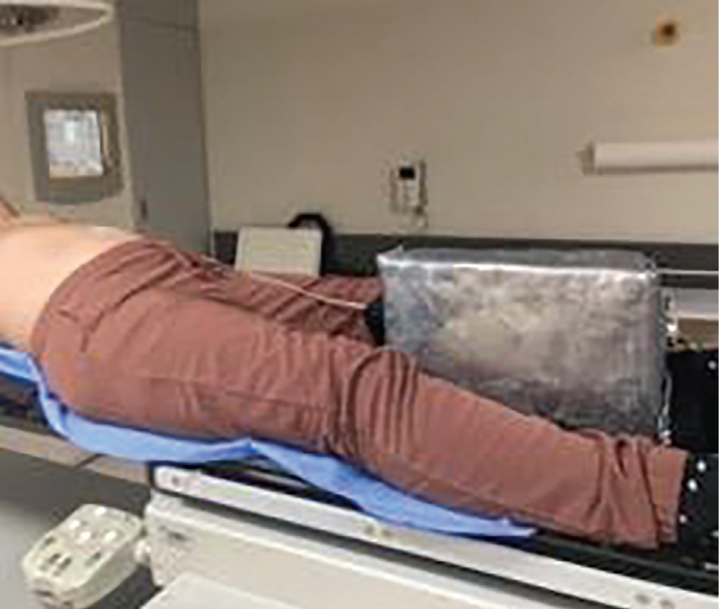
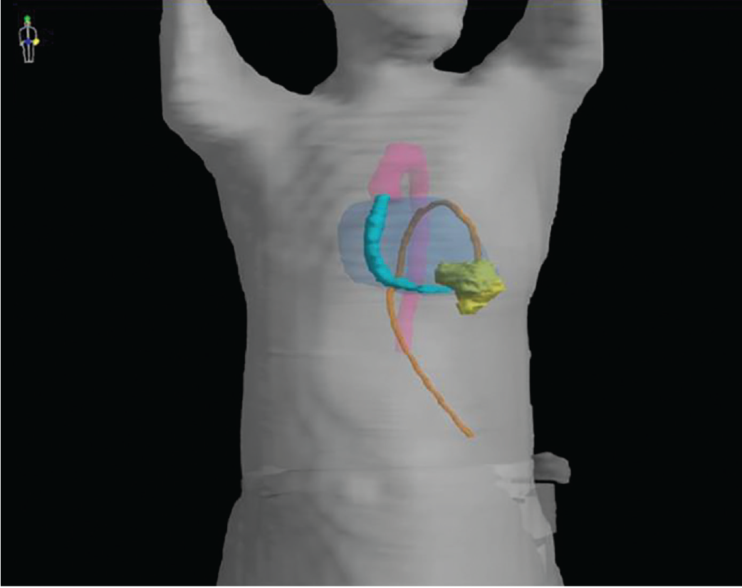
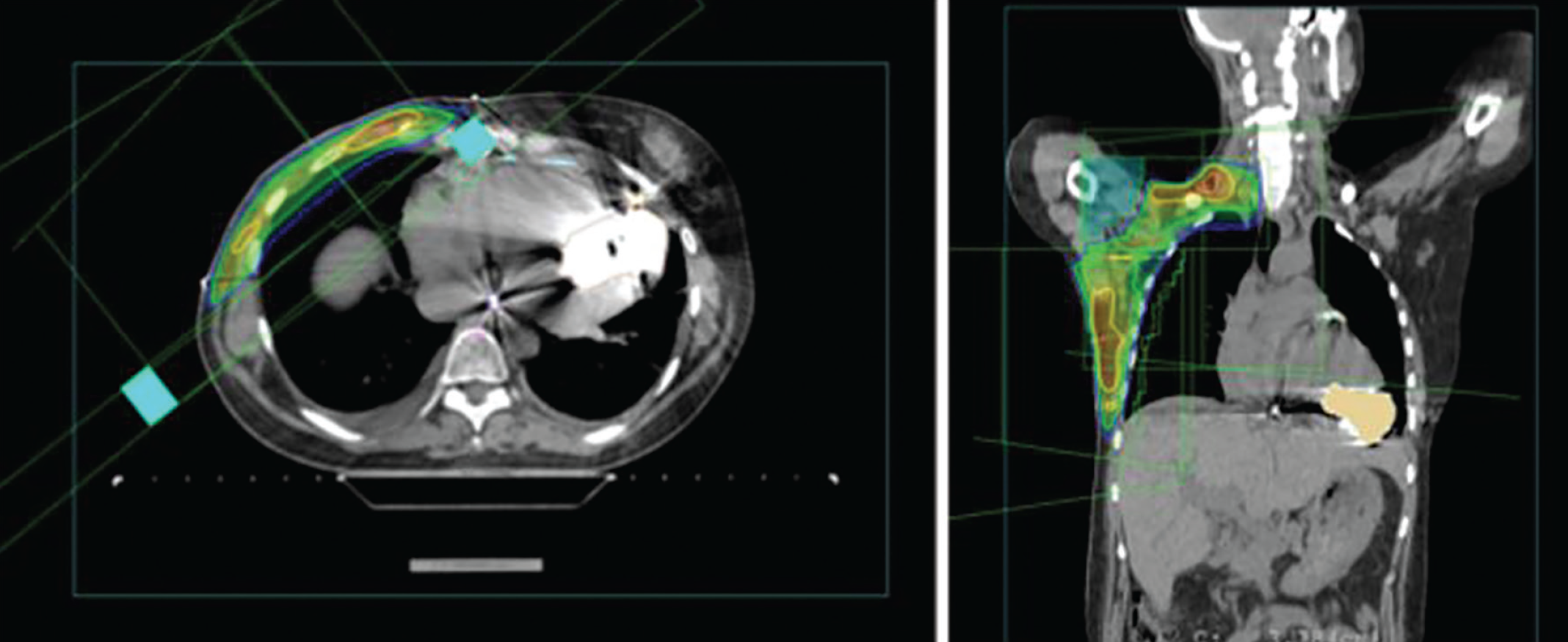
External LVAD parts were carefully observed, and a 5 mm-thick lead shield box for the external system controller was produced. The patient was placed on a breast board with arms above the head (). Radiotherapy was prescribed to the right chest wall and regional lymphatics (axillary levels 1–4) for 50 Gy in 25 fractions. Intracorporeal LVAD parts were delineated separately (). An anterior supraclavicular field and opposed 6 MV photons to the right chest wall area () were used for treatment to minimize radiation exposure to LVAD subparts. Radiation doses received by separate parts of LVAD is shown in . A cardiology nurse and device specialist were available for each treatment and conducted daily pre- and post-treatment measurements of flow and power as a surrogate for LVAD performance, which showed no major measurable alterations. Treatment was completed without any device errors or malfunctions (). During cardiologic follow-up visits, no signs of LVAD malfunction were observed.
LVAD: Left ventricular assist devices
LVAD: Left ventricular assist devicesac
Table 1. Radiation doses received by different parts of LVAD
- | Maximum dose (cGy) | Mean dose (cGy) |
LVAD pump | 24 | 9 |
Outflow graft | 767 | 227 |
Driveline | 199 | 52 |
LVAD: Left ventricular assist devices
Table 2. Measured device flow and power parameters during treatment
- | Flow (L/min) | Power (watt) |
|---|---|---|
1st Fraction - beginning | 3.3 | 3.2 |
6th Fraction - week 2 | 3.9 | 3.4 |
11th Fraction - week 3 | 3.7 | 3.4 |
16th Fraction - week 4 | 3.9 | 3.4 |
21st Fraction - week 5 | 4.2 | 3.6 |
25th (Last) fraction | 3.3 | 3.2 |
Oncological follow-up continued with no local or systemic breast cancer recurrence. Forty-nine months after breast cancer diagnosis and 41 months after LVAD placement, she had a non-traumatic intracranial hemorrhage and underwent decompression surgery, and unfortunately, died due to sepsis during post-op care.
Discussion and Conclusion
Deciding whether to give radiotherapy for patients with LVAD may be challenging, considering the scarcity of high-quality data. Herein, we describe a breast cancer patient with LVAD who safely received adjuvant radiotherapy. Accumulated doses caused no disturbance to LVAD function during follow-up. To the best of our knowledge, this is the first patient who had a HeartWare LVAD and received adjuvant radiation therapy for primary breast cancer.
In terms of the safety of radiotherapy with LVADs, a review by Spano et al. (), reported that LVAD performance was unaffected for doses of up to 70-75 Gy which are considered in therapeutic range for both photon and proton beams in several in vitro studies. However, Sindhu et al. () found, while the pump components were resilient to the 70 Gy of proton irradiation, the driveline part showed functional disturbance at 30 Gy of continuous proton irradiation, necessitating careful evaluation of various parts of the LVAD in dose-volume analysis.
To date, only twelve case reports, including eighteen cases, have been published (, -). These reports evaluated radiotherapy safety in various tumor sites and with different LVAD brands [HeartMate, Abbott, Chicago, IL, USA (HM II-III)]; HeartWare, HeartWare Inc., Miami Lakes, FL (HW); Thoratec, Thoratec, Pleasanton, CA; Novacor LVAD Atlas II VR SN, St. Jude Medical, Saint Paul, Minnesota, US. In most studies, patients were implanted with a HeartMate LVAD.
Eight studies evaluated nine patients treated with conventionally fractionated radiotherapy, while six studies evaluated nine patients treated with stereotactic body radiotherapy (SBRT) (, , , , ). One study reported a lung cancer patient treated with proton beams (). Studies reporting cardiac ablative therapies with SBRT for arrhythmias were reviewed by Benali et al. (), with no LVAD-related complications after SBRT and are not included in this review. In fifteen cases, radiotherapy fields involved the thoracic region, in two cases, encompassed pelvic areas, and in one case, treatment was directed to the whole body.
In the studies reporting the variables that reflect the performance of the LVAD, such as power, flow, and rotational speed, all report insignificant changes between pre- and post-irradiation values (, , ). None of the studies reported any disturbance in device performance.
Previously published case reports are summarized in (, -). Most of the studies used 6 to 15 MV photon beams with prescribed doses within the range of 20–66 Gy delivered in 3–33 fractions. Mean doses received by LVAD ranged between 8–1922 cGy, and maximum doses to LVAD and its subparts varied according to laterality of the tumor up to 6830 cGy (). There was no reported device malfunctioning, with the longest reported follow-up being 29 months. We believe that this report will contribute to the current literature in several aspects, notably by reporting the successful delivery of radiotherapy in the first case with breast cancer primary and the second case with an implanted HeartWare LVAD. Furthermore, this report also reports the longest follow-up with 41 months and no device malformation.
Table 3. List of the studies reporting patients irradiated with VAD for various sites and techniques
Study | Year | Device | Diagnosis | Beam | Prescription dose (cGy) | Fraction | Max dose (cGy) to the device | Mean dose (cGy) to the device |
|---|---|---|---|---|---|---|---|---|
Lasher et al. () | 2008 | Thoratec | Rectal adenocarcinoma | 15 MV photon | 4500 | 25 | 425 | - |
Netuka et al. () | 2013 | HM II | Hodgkin lymphoma | NR | NR | 12 | NR | NR |
Scobioala et al. () | 2015 | Novacor LVAD Atlas II VR SN (St. Jude Medical, Saint Paul, Minnesota, US) | Lung squamous cell carcinoma | Conventional: 6/15 MV photon beams | 2520 | 14 | 538 | 231 |
SBRT: 6 MV | 3500 | 5 | ||||||
Emerson et al. () | 2016 | HM II | Gastroesophageal junction adenocarcinoma | 15 MV photon | 5040 | NR | 4900 | 1922 |
- | 2016 | HM II | Lung adenocarcinoma | SBRT: 6 MV photon | 5400 | 3 | 61 | 9.6 |
- | 2016 | HM II | Lung cancer/vertebral mets | 6/15 MV photon | 2000/3000 | NR | 2450 | 1423 |
Ostertag-Hill et al. () | 2018 | HeartWare | Lung nodule | SBRT: 6 MV photon | 5000 | 5 | 698 | 45 |
Spano et al. () | 2019 | HM III | Lung adenocarcinoma | SBRT: 6 MV photon | 5000 | 5 | VAD: 29 | VAD: 8 |
Outflow graft: 991 | Outflow graft: 147 | |||||||
Drive line: 34 | Drive line: 11 | |||||||
Sato et al. () | 2020 | NR | Thymic carcinoma | 6 MV photon | 6600 | 33 | Outflow graft: 6830 | - |
VAD: 0 | - | |||||||
CRT-D: 99 | - | |||||||
Schumer et al. () | 2022 | HM II | Lung squamous cell carcinoma | Proton beams | 6000 | 30 | NR | NR |
Yousafzai et al. () | 2022 | HM III | Sternal osteosarcoma | NR | 6660 | NR | 46.4 | - |
Butt and Sheikh () | 2023 | HM III | Lung adenocarcinoma | SBRT | 3000 | 5 | NR | NR |
- | 2023 | HMIII | Lung small cell carcinoma | SBRT | 5000 | 5 | NR | NR |
Webster et al. () | 2024 | HM III | Acute myeloblastic leukemia | 16 MV photon | 400 | 2 | 120 | - |
Hayashi et al. () | 2024 | - | Lung adenocarcinoma | SBRT | 42-52 Gy | 4 | <200 | - |
Lung adenocarcinoma | SBRT | 42-52 Gy | 4 | <200 | - | |||
Lung adenocarcinoma | SBRT | 42-52 Gy | 4 | <200 | - | |||
Alanyalı et al. | 2025 | HeartWare | Breast invasive ductal carcinoma | 6 MV photon | 5000 | 25 | Outflow graft: 767 | Outflow graft: 227 |
VAD: 24 | VAD: 9 |
NR: Not reported; LVAD: Left ventricular assist devices
With both in vitro and in vivo results stating the safe irradiation of LVAD and the review by Spano et al. () summarizing the recommendations, including ensuring a multidisciplinary approach, using beam energies <10 MV to minimize neutron contamination, ensuring a rapid response team is available, close monitoring of the patient, securing the extracorporeal parts, and interrogating the LVAD after each radiation session. In the presented case, contralaterality of the LVAD and primary tumor location made reducing radiation dose received by LVAD relatively simple, but in cases with close proximity between tumor site and the device, the radiotherapy becomes significantly more complex. Direct irradiation of the device may be unavoidable. In such situations, careful multidisciplinary planning is essential to balance optimal oncologic outcomes with device safety. Whenever feasible, attempts to reduce the dose received by the LVAD should be made, including advanced radiotherapy techniques such as intensity modulated radiotherapy or proton irradiation, or distancing the device from the radiotherapy field with techniques like deep inspiration breath hold.
Cancer patients with LVAD in situ pose a multifaceted challenge in radiotherapy in terms of treatment decision and technical considerations. The presented case described a patient who received adjuvant radiotherapy to the thoracic region, and yielded no evidence of device malfunction, thereby affirming its safe implementation in this case. There is limited published data regarding the safety of radiotherapy in patients with LVAD. More studies are needed in this area to ensure optimal patient safety and treatment decision-making.
Footnotes
Ethics
Informed Consent: Written informed consent was taken from the patient.
Authorship Contributions
Surgical and Medical Practices: S.A., E.K., M.K., Ü.K., S.N.; Concept: S.A., E.K., Ü.K., S.N.; Design: S.A., E.K., M.K., Ü.K., S.N.; Data Collection or Processing: S.A., E.K., S.N.; Analysis or Interpretation: S.A., E.K., S.N.; Literature Search: S.A., E.K.; Writing: S.A., E.K.
Conflict of Interest: No conflict of interest was declared by the authors.
Financial Disclosure: The authors declared that this study received no financial support.
References
Molina EJ, Shah P, Kiernan MS, Cornwell WK 3rd, Copeland H, Takeda K, et al. The Society of Thoracic Surgeons Intermacs 2020 annual report. Ann Thorac Surg. 2021; 111: 778-792. (PMID: 33465365)
Spano G, Stutz E, Elicin O, Hugi B, Henzen D, Fürholz M, et al. Is it safe to irradiate the newest generation of ventricular assist devices? A case report and systematic literature review. Artif Organs. 2020; 44: 449-456. (PMID: 31769042)
Sindhu KK, Shi C, Moss N, Lin H, Zhang J, Hu L, et al. The effects of pencil beam scanning proton beam therapy on a heartmate 3 left ventricular assist device: implications for patient safety. ASAIO J. 2022; 68: e145-e147. (PMID: 35537112)
Benali K, Lloyd MS, Petrosyan A, Rigal L, Quivrin M, Bessieres I, et al. Cardiac stereotactic radiation therapy for refractory ventricular arrhythmias in patients with left ventricular assist devices. J Cardiovasc Electrophysiol. 2024; 35: 206-213. (PMID: 38018417)
Lasher DE, Wojcicka JB, Malcom R, Shears LL. Case study of radiation therapy treatment of a patient with a cardiac ventricular assist device. J Appl Clin Med Phys. 2008; 9: 214-220. (PMID: 19020490)
Netuka I, Stepankova P, Urban M, Maly J, Szarszoi O, Dorazilova Z, et al. Is severe cardiac dysfunction a contraindication for complex combined oncotherapy of Hodgkin’s lymphoma? Not any more. ASAIO J. 2013; 59: 320-321. (PMID: 23644622)
Scobioala S, Ernst I, Moustakis C, Haverkamp U, Martens S, Eich HT. A case of radiotherapy for an advanced bronchial carcinoma patient with implanted cardiac rhythm machines as well as heart assist device. Radiat Oncol. 2015; 10: 78. (PMID: 25885061)
Emerson LY, Deek MP, Almendral J, Jabbour SK. Radiation therapy in patients with left ventricular assist device: a case report and literature review. Pract Radiat Oncol. 2016; 6: e145-e147. (PMID: 27017262)
Ostertag-Hill CA, Mudd J, Werle DP, Tieu BH, Nabavizadeh N. Safe delivery of lung stereotactic body radiation therapy in a patient with a left ventricular assist device and implantable cardioverter defibrillator. Clin Case Rep. 2018; 6: 1704-1707. (PMID: 30214746)
Sato GE, Matsuo Y, Kanemitsu H, Minatoya K, Nakajima D, Date H, et al. Safe delivery of postoperative radiotherapy for thymic carcinoma located on the outflow graft of a left ventricular assist device. JTO Clin Res Rep. 2020; 2: 100101. (PMID: 34589971)
Schumer EM, Schettle S, Stulak JM, Rosenbaum AN. First report of proton beam therapy in a patient with a left ventricular assist device. ASAIO J. 2022; 68: e84-e86. (PMID: 35503645)
Yousafzai OK, Stewart SM, Zaman MO, Ingenito A, Kim B. Sternal radiation for osteosarcoma treatment in a patient with left ventricular assist device. JACC. 2022; 79 (9_Supplement): 2579.
Butt N, Sheikh FH. Feasibility of mediastinal radiation use in heartmate 3 LVAD recipients with cancer. Methodist Debakey Cardiovasc J. 2023; 19: 15-19. (PMID: 36742441)
Webster M, Dona Lemus OM, Zheng D, Wancura JN, Tanny S, Sakthivel G, et al. Case study with dosimetric analysis: total body irradiation to a patient with a left ventricular assist device. Clin Case Rep. 2024; 12: e8868. (PMID: 38756618)
Hayashi K, Ishikawa H, Fujiwara K, Nakai M, Ono H, Fumimoto Y, et al. Stereotactic ablative radiotherapy for early-stage lung cancer in patients with left ventricular assist device: a case series. Anticancer Res. 2024; 44: 4113-4117. (PMID: 39197891)