
Abstract
Dermatofibrosarcoma protuberans (DFSP) is a rare, low-grade, fibroblastic mesenchymal tumor derived from the dermis. Breast is an uncommon site with an incidence of only 0.8–4.5% and an overall population incidence at any site of 4.2–4.5 per million. Surgical excision with 2–3 cm margin is the gold standard treatment. Selected cases are subjected to radiotherapy or systemic therapy with Imatinib. Due to the rare presentation, we report a similar case of DFSP on the left breast in a 42-year-old woman, who was initially diagnosed with benign phyllodes tumor of the left breast and final histopathology report of the wide local excision specimen diagnosed DFSP of the breast.
Keywords: Breast, cutaneous lesion, dermatofibrosarcoma protuberans, keloid-like
Key Point
• Dermatofibrosarcoma protuberans on breast is an unusual location of the tumor, which can be confused with phyllodes due to its rarity.
Introduction
Dermatofibrosarcoma protuberans (DFSP) is a rare, low-grade, fibroblastic mesenchymal tumor originating from the dermis. It most commonly affects the trunk, followed by the proximal extremities (). Involvement of the breast is uncommon, with an estimated incidence of 0.8–4.5% among DFSP cases, and an overall DFSP incidence of only 4.2–4.5 per million population (-). Surgical excision with 2-3 cm margin is the gold standard treatment (, ). Selected cases are subjected to radiotherapy or systemic therapy with Imatinib ().
Due to its rare presentation, breast DFSPs are often misdiagnosed during clinical examination, commonly mistaken for dermatofibroma, hemangioma or fibroepithelial lesions, as clinicians are often not familiar with its occurrence in the breast ().
Here, we report a case involving a 42-year-old woman with DFSP of the left breast, which was initially diagnosed as a benign phyllodes tumor based on core needle biopsy. However, the final histopathological evaluation of the wide local excision specimen diagnosed DFSP of the breast.
Case Presentation
A 42-year-old woman presented with a discolored protruding lesion on the skin of the left breast which was insidious in onset and gradually progressive over the course of two years from approximately 2x1 cm to around 4x3 cm at presentation. The lesion was painless and non-itchy. She did not have any contributory family history. On clinical examination, a lobulated, exophytic, keloid-like lesion with pink to whitish discoloration was noted on the left breast, located just above the inframammary fold, extending from the 6 to 5 o’clock position. The lesion was well-defined, measured 4.2×3.0 cm, and was fixed to the overlying skin but free from the underlying breast tissue (). The right breast, the remaining left breast and bilateral axillae were unremarkable.
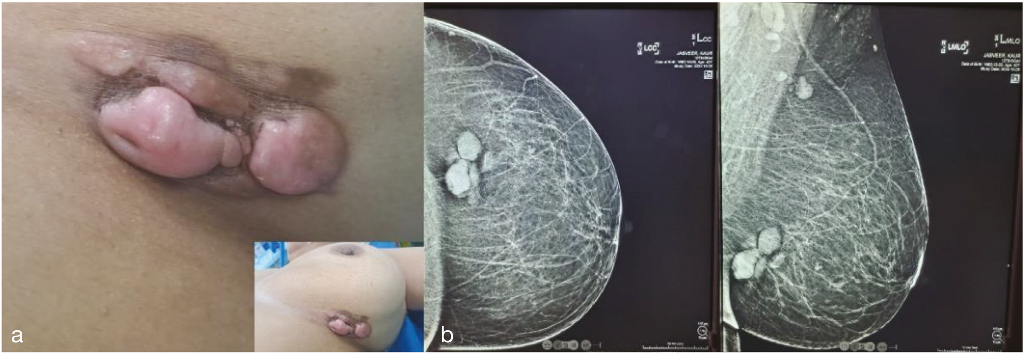
A mammogram with ultrasound correlation was performed. Mammogram () demonstrated two high-density, lobulated lesions with well-circumscribed margins in the lower outer quadrant of the breast,12 cm away from the nipple measuring 2.6x2.1 cm and 2x1.9 cm respectively. No intramammary lesion or microcalcification were observed. Ultrasound correlation revealed a few well defined, oval shaped, solid hypoechoic lesions, the largest measuring 3x1.3 cm located at 5 o’clock position in the left inframammary fold, about 10 cm away from the nipple/areolar complex. The lesion exhibited cleft-like cystic spaces with posterior acoustic enhancement and significant internal vascularity on color Doppler. Axillae were normal. The findings were suggestive of phyllodes tumor. For histopathological confirmation, a core needle biopsy was done which showed a mesenchymal tumor, favoring benign phyllodes. It had ovoid to spindled shaped cells with no epithelial component. So, as this was a breast lesion with such morphology, phyllodes is the usual diagnosis ().
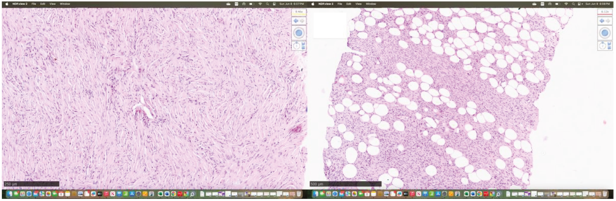
H&E: Hematoxylin and eosin
Hence, no immunohistochemistry (IHC) was planned initially. The patient subsequently underwent wide local excision with 2 cm margin.
The perioperative period was uneventful. On histology, the resected specimen showed dermal-based spindle cell tumor with infiltration into the subcutaneous fat, sparing the underlying breast parenchyma. This tumor was composed of uniform, bland, spindle cells arranged in a storiform pattern with areas of whorled fascicles and characteristic entrapment of adnexal structures. No significant mitosis, nuclear atypia or necrosis were seen. On IHC these cells were diffusely positive for CD34 (, ). Features were consistent with DFSP. For confirmation, fluorescence in situ hybridization was done on the paraffin embedded tissue block, which showed PDGFB gene rearrangement ().
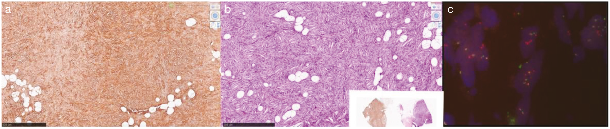
H&E: Hematoxylin and eosin
It is important to note that diagnosing DFSP with small core biopsy samples can be challenging, particularly in unusual anatomical locations such as the breast. In such limited samples where the representation of tumor is not adequate, the potential misdiagnosis with more common entities like benign phyllodes, solitary fibrous tumor, fibromatosis and cellular fibrous histocytoma is common. Given these challenges, a carefully selected IHC panel including CD34, STAT6, Beta catenin, S100 and Ki-67 is essential to distinguish DFSP from its closest morphological mimics in small biopsy samples. Informed consent was taken from patient.
Follow-up
The patient was planned for close follow-up post surgery. Clinical examination at three months of follow-up showed no evidence of recurrence and the surgical scar was healthy. A follow-up mammogram performed subsequently (post 3-months) showed no residual lesion or new abnormalities, indicating a favorable postoperative course.
Footnotes
Ethics
Informed Consent: Informed consent was taken from patient.
Authorship Contributions
Surgical and Medical Practices: R.M., R.M., B.K.S.; Concept: R.M., R.M., B.K.S.; Design: R.M., R.M., B.K.S.; Data Collection or Processing: R.M., R.M., B.K.S.; Analysis or Interpretation: R.M., R.M., B.K.S.; Literature Search: R.M., R.M., B.K.S.; Writing: R.M., R.M., B.K.S.
Conflict of Interest: No conflict of interest was declared by the authors.
Financial Disclosure: The authors declared that this study received no financial support.
References
Wang Y, Wang Y, Chen R, Tang Z, Liu S. A rare malignant disease, dermatofibrosarcoma protuberans of the breast: a retrospective analysis and review of literature. Biomed Res Int. 2020; 2020: 8852182. (PMID: 33224981)
Ramesh O, Leila Haji M, Sadaf A. Corrigendum to “dermatofibrosarcoma protuberance of the skin of the breast: a case study and review of the literature” [Int J Surg Open 50 (2023) 100583]. IJS Open 52: 100591, March 2023.
Dimas D, Boutas I, Potiris A, Koufopoulos N, Balalis D, Sitara K, et al. Dermatofibrosarcoma protuberans of the breast: a case study. Mol Clin Oncol. 2021; 14: 50. (PMID: 33604040)